Research
PET and MRI for Brain Imaging
- Low-dose PET Image Reconstruction using Deep Learning
- 4D PET Image Reconstruction using Deep Learning
- PET Tau and Amyloid-β Imaging to Study Alzheimer’s Disease
- Imaging Markers for Neurodegenerative Diseases
In this figure, [18F]FDG brain images are shown for two different reconstruction methods: Maximum Likelihood Expectation Maximization (MLEM) algorithm, the standard PET image reconstruction method, and MR-Assisted MAP-EM reconstruction. The animation shows: 1) the same slice converging to a solution during the reconstruction process, 2) all the slices for the final reconstructed image, and 3) rotating Maximum Intensity Projections (MIPs).
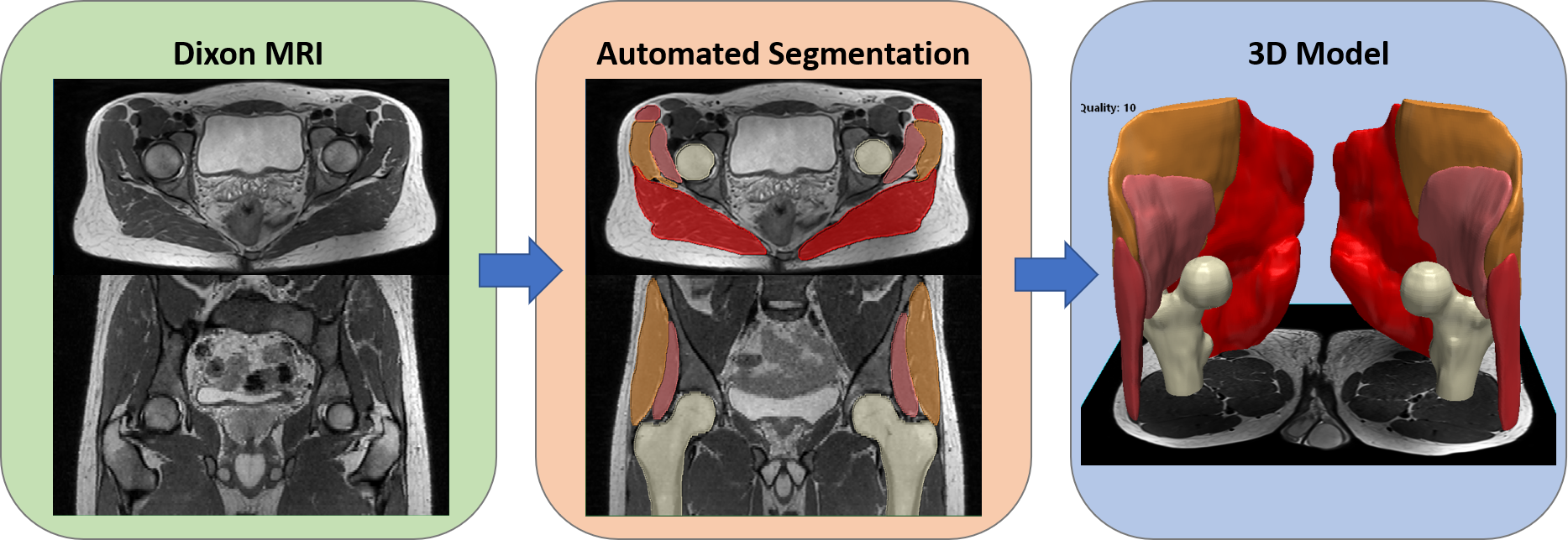
MRI and CT for Musculoskeletal Applications
- Deep Learning Segmentation Methods for Musculoskeletal Applications
- MRI Biomarkers to Study Sarcopenia and Muscle Health
- Deep Learning for Metal-artefacts Reduction Methods